HUMAN REMAINS
| MIDDLE BRONZE AGE HUMAN REMAINS FROM TELL ARBID (SECTOR P) |
Dr. Arkadiusz Sołtysiak/University of Warsaw
INTRODUCTION
During archaeological excavations at Tell Arbid, skeletal remains of at least 311 individuals were found, mainly dated to the 3rd and 2nd millennia BCE (Sołtysiak 2010a). From this number, at least 92 skeletons were excavated between 2008 and 2010 in Sector P. A preliminary report concerning this assemblage (Sołtysiak & Koliński 2011) and a case study on one individual (Pitre et al. 2017) have been published elsewhere, and here a brief catalogue of the Middle Bronze Age (MBA) human remains (see Table 1) is provided together with information about data recording protocol. The full dataset is available from the author upon request.
METHODS
All human remains were studied at the dig house, and therefore a simplified set of measurements and observations has been recorded, based on the protocol by Buikstra and Ubelaker (1994). Available measuring tools were spreading caliper, sliding caliper (GPM Swiss), osteometric board (made from three plywood boards and a millimeter paper, verified with the spreading caliper), protractor and a strap of paper for measuring circumferences. Taphonomical observations were not scored in a systematic way, but unusual weathering (Behrensmeyer 1978), staining, root etching or insect tunneling (Pittoni 2009) were recorded together with general remarks on bone fragmentation pattern and possible specific pattern of postmortemdamage (Sołtysiak 2010b).
Completeness pattern was scored on a 4-point scale (0 – absent element, 1 – less than 50% present, 2 – more than 50% present, 3 – complete or nearly complete element). The list included major skull bones (left and right side scored separately), i.e. frontal, parietal, occipital, temporal, sphenoid, zygomatic, nasal, maxilla, palatine and mandible, six long bones scored separately for five areas (proximal end, proximal 1/3 of the shaft, midshaft, distal 1/3 of the shaft, distal end), six parts of the spine (atlas, axis, other cervical, thoracic, lumbar and sacral vertebrae), ribs (irrespective side), sternal manubrium and sternal body, hyoid, hand and foot bones (irrespective side) and the following elements with left and right side scored separately: clavicle, scapula, ilium, ischium, pubis, talus, calcaneus, patella. Additionally, the presence of minor bones (carpals, metacarpals, tarsals, metatarsals and phalanges) was noted without a completeness score.
Sex assessment was based primarily on the morphology of os coxae, including ventral arc, subpubic concavity, ischiopubic ramus ridge (Phenice 1969), greater sciatic notch and preauricular sulcus. For skull, the robustness of the following landmarks was recorded: nuchal crest, mastoid process, supraorbital margin, glabella, mental eminence. Age-at-death assessment was based on the morphology of pubic symphysis, scored using the protocols by Todd (1920) and Brooks and Suchey (1990), and of the auricular surface (Meindl & Lovejoy 1989). For bilateral morphological landmarks, both sides were scored separately. Additionally, suture closure at the external cranial vault (midlamboid, lambda, obelion, anterior sagittal, bregma, midcoronal, pterion, sphenofrontal, inferior and superior sphenotemporal), palate (incisive, anterior and posterior median palate, transverse palate) and internal cranial vault (sagittal, left lambdoid, left coronal) were scored on the 4-point scale (Meindl & Lovejoy 1985).
The set of 24 standard cranial measurements includes maximum cranial length, maximum cranial breadth, bizygomatic diameter, basion-bregma height, cranial base length, basion-prosthion length, maxillo-alveolar breadth and length, biauricular breadth, upper facial height and breadth, minimum frontal breadth, nasal height and breadth, orbital breadth and height, biorbital breadth, interorbital breadth, frontal, parietal and occipital chords, foramen magnum length and breadth, mastoid length (Buikstra & Ubelaker 1994). Orbital breadth was measured in two ways (dakryon to ectoconchion and maxillofrontale to ectoconchion, cf. Nikita 2017). Ten standard measurements of the mandible include chin height, height of the mandibular body, breadth of the mandibular body, bigonial width, bicondylar breadth, minimum ramus breadth, maximum ramus breadth, maximum ramus height, mandibular length, mandibular angle (Buikstra & Ubelaker 1994). Additionally, gonion-gnathion length, condylo-symphyseal length and maximum length of the condyle were measured.
The list of postcranial measurements included all 44 standard measurements defined in Buikstra & Ubelaker (1994), with the following additions: glenoid height (scapula), maximum and minimum measurement at the maximum development of the deltoid tuberosity (humerus), maximum proximal and distal epiphyseal breadths, antero-posterior and medio-lateral diameter at the maximum development of the interosseous crest (radius), maximum breadth at the coronoid process (ulna), maximum length of the sacrum, maximum diameter of the acetabulum, maximum length of tibia, maximum proximal and distal epiphyseal breadths (fibula), breadth and length of atlas, maximum height, height without the odontoid process, breadth and length of axis, height and breadth of patella, total length and maximum length of upper articular facet (talus), breadth of the navicular, maximum height and maximum breadth of the sternal body (without xyphoid process), maximum height and maximum breadth of the sternal manubrium, maximum length, proximal and distal epiphyseal breadths, antero-posterior and medio-lateral diameters at midshaft of the first metacarpal. All measurements were taken on the left side or on the right side if the left one was missing. Only the measurements of the first metacarpal were taken on both sides.
The standard list of 36 primary nonmetric traits (Buikstra & Ubelaker 1994) was supplemented by the division of talar articular surface on calcaneus, third trochanter and lateral fossa on proximal femur, supracondylar ridge on distal humerus, vastus notch on patella, squatting facet on distal tibia and spina bifida on sacrum. Pathological conditions were documented with photographs, and some specific conditions, such as cribra orbitaliaand porotic hyperostosis (if more than 75% of cranial vault was present), were scored in a simplified way (Steckel et al.2006). Degenerative joint disease in all joints of which more than 75% was preserved was assessed on a 3-point scale (Steckel et al.2006).
For every tooth, the following data were gathered: mediodistal and buccolingual maximum diameter, dental wear score on a 10-point scale (molars, Smith 1984), minimum and maximum score for four quadrants, or 8-point scale (other teeth, Scott 1979), enamel hypoplasia and dental caries. Enamel hypoplasia was scored on a 4-point scale (0 – no hypoplasia, 1 – visible lines that are not perceptible with a fingernail, 2 – one line perceptible with a fingernail, 3 – two or more perceptible lines, Steckel et al.2006, modified) and dental caries was scored on a 4-point scale (0 – no lesions, 1 – a lesion less than 2 mm in breadth, 2 – a lesion 2–6 mm in breadth, 3 – a larger lesion, Sołtysiak 2014). Apart from this, the location of the lesion was noted (medial, distal, labial, lingual, occlusal; root, cemento-enamel junction, crown). Also, antemortemand postmortemtooth loss were noted as well as absorption of the alveolar process >2 mm. Presence of a considerable amount of dental calculus was also observed but with no specific quantitative scale.
CATALOGUE OF HUMAN REMAINS
GP02 (35/60 G4). A fairly complete and relatively well-preserved skeleton of a 3-year-old child. Bones were slightly weathered, with spots of crystalline deposits. Very clear linear enamel hypoplasia has been observed at all developing crowns of anterior permanent teeth. Initial cribra orbitaliawas present in the left orbita.
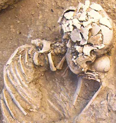
GP03 (35/60 G3). A fairly complete skeleton of a 9-month-old infant with evidence of weathering (Fig. 1 Evidence of weathering on a cranial vault fragment.), spots of crystalline deposits and many elements fragmented to small pieces. There were also some elements from a subadult animal. The left clavicle was deformed near the conoid tubercle (Fig. 2 Deformation of the left clavicle with some crystalline deposit.).
GP04 (35/60 G5). A fairly complete and relatively well-preserved skeleton of a 3-year-old child. Bones were slightly weathered and mixed with remains of a subadult animal. Initial cribra orbitaliawas accompanied by hypervascularization and endocranial porosity around the sagittal sulcus of the occipital bone (Fig. 3 Hypervascularization and endocranial porosity on the occipital bone.). Very clear linear enamel hypoplasia has been observed at developing crowns of anterior permanent teeth.
GP05 (35-60 G6). A complete and well-preserved skeleton of an adult female. Both tibial proximal epiphyses sloped down backwards at an angle of 15–20º. The right humerus was much shorter than the left one, with a dislocated articular surface (Fig. 4 Asymmetry of humeri.). This deformation was likely a congenital condition and did not affect forearm bones except for some asymmetry in the supinator crest of the ulna. The skull was almost complete (Fig. 5 Lateral view of the cranium.).
GP06 (35-61 G1). A skeleton of a female in an average state of preservation and with occasional large crystalline deposits on the bone surface. The cranium is missing. The fifth lumbar and first sacral vertebrae were fused with large osteophytes (up to 15 mm) on the upper margin of the L5 body. Also the sternal manubrium and body were fused.
GP07 (35/61 G2). A multiple burial with mixed elements from adult skeletons. Most of them were strongly eroded. The minimum number of individuals was based on the number of tali and first coccygeal vertebrae. The impression of the costoclavicular ligament on the relatively well-preserved right clavicle is extremely deep and broad (Fig. 6 Impression of the costoclavicular ligement on the right clavicle.). The sternal epiphysis of the clavicle and annular rings on some vertebrae were not fused, indicating a young age of at least one individual.
GP10 (37/60 G2). At least two poorly preserved disarticulated skeletons (one adult and one subadult individual). The adult skeleton was roughly complete, and the subadult one was represented by a few bone fragments from both upper and lower extremity as well as most of the teeth.
GP12 (37/60 G4). A roughly complete but poorly preserved skeleton of a likely female individual, affected by insect tunneling. One toe phalanx was fractured and well healed (Fig. 9 Healed fracture of a foot phalanx.). The alveolar process was reduced by 3–4 mm, and several molars were affected by dental caries.
GP13 (37/60 G5). An almost complete and relatively well-preserved skeleton of an adult female individual, with only small fragments of the cranium. Bones were slightly weathered and covered by large crystalline deposits; occasionally also root etching was present. Muscular attachment areas were not well developed, but a short, broad and bifurcate supracondylar spur was present on the humerus (Fig. 10 Supracondylar spur on humerus.). Many carious lesions were present on molar teeth.
GP17 (37/59 G3). A very poorly-preserved but fairly complete skeleton of a 9-month-old infant, with evidence of weathering and root etching (Fig. 7 Evidence of root etching on a cranial vault fragment.). A single deciduous incisor of another individual was also present in this assemblage.
GP18 (37/59 G4). An incomplete and heavily fragmented skeleton of an adult individual, likely female. The cortical bone in the shafts of the long bones was very thin, indicating osteoporosis. Several joints were affected by advanced degenerative joint disease. A healed fracture in the distal part of the midshaft is attested on one small fragment. The cranial vault was relatively thick, up to 9 mm, with broad diploë. All anterior mandibular teeth were lost ante-mortem. More details about this individual can be found in Pitre et al.2017.
GP19 (37/59 G5). A poorly preserved and incomplete skeleton of a gracile adult individual, likely female. Weathering and root etching were common (Fig. 8 Evidence of extreme weathering and erosion on femoral midshaft.).
GP21 (37/61 G2). An almost complete and relatively well-preserved skeleton of a foetus (8 months in utero).
GP22 (37/62 G1). A small assemblage of eroded bones and many teeth of at least four individuals including one neonate and three children 4–5 years old.
GP23 (37/62 G2). An incomplete and poorly preserved skeleton of a child, with slight evidence of weathering. Not only was enamel hypoplasia present on the germs of permanent teeth but an irregular hypoplastic area-type lesion was also observed on the left deciduous canine.
GP25 (37/62 G4). Mixed strongly eroded elements from an adult individual and six children of different age-at-death. Some of them were also affected by root etching. Most individuals were represented only by teeth and/or a few bone fragments; many animal elements were also present. Anterior dentition of a 10-year-old child shows extensive evidence of linear enamel hypoplasia.
GP26 (37/62 G7). A multiple deposit of adult and subadult skeletons; many animal elements were also present. Bones were strongly eroded, and many individuals were represented only by a few teeth/bone fragments.
GP27 (37/62 G6). An incomplete and poorly preserved skeleton of a 2-year-old child together with several bone fragments of a subadult animal. Weathering was evident on some elements (Fig. 12 Evidence of weathering on ischium.).
GP28 (37/62 G5). An eroded but fairly complete skeleton of a 2-year-old child, mixed with many elements from a subadult animal. Most elements from the left upper extremity and many parts of the skull were missing. An initial phase of cribra orbitaliahas been observed in the left orbita (Fig. 11 Cribra orbitalia.).
GP29 (37/62 G8). A few elements of an infant in a secondary deposit.
GP31 (37/62 G9). A very small bone assemblage with remains of at least two individuals, one adult and one 2-year-old infant.
GP32 (37/63 G1). Very eroded and weathered bone fragments of four individuals. All skeletons were highly incomplete, and the attribution of elements to specific individuals was based only on archaeological documentation. It was likely a secondary deposit, as the colour of teeth attributed to two individuals was different (light beige and brownish respectively). The cranial vaults were highly incomplete, but on one of them, an area of obliterated porosity can be observed (Fig. 13 Porosity on a cranial vault fragment.).
GP34 (38/61 G1). Bone fragments of at least three individuals: two adults (identified based on the teeth; one mandible was relatively robust, suggesting male sex) and one child. The skeletons were incomplete and poorly preserved, sometimes affected by root etching.
GP51 (38/61 G14). Two very incomplete and poorly preserved skeletons of subadult individuals (one neonate and one c. 3-year-old child). Originally, they may have been complete and articulated, but the degree of erosion is high in this case, and some elements were affected by root etching and insect tunneling. Moderate cribra orbitaliaand initial porotic hyperostosis over lambda can be seen on the cranium of the older individual.
GP56 (36/62 G4). A strongly eroded skeleton of an adult female. The left femur had likely been broken in the distal part of the shaft and healed completely with some dislocation, but only a small fragment of this area is preserved. Clear asymmetry was observed in long bones of the upper extremity, and especially in the prominence of the supinator crest in the ulna. Spondylosis in all preserved cervical vertebrae, degenerative joint disease in the distal femur and reduction of the alveolar process by 6 mm are likely the consequence of the advanced age-at-death of this individual.
GP57 (36/62 G5). A very small number of human elements retrieved from an assemblage of animal bones. One adult individual was represented only by a fragment of a patella and distal epiphysis of a metacarpal. There are also a few bone fragments of children and dentition of at least three individuals with roughly the same age-at-death of c. 3 years (MNI based on the number of left mandibular first molars). Their remains were likely moved to the secondary deposit from different places, as the colour of teeth attributed to specific individuals is different: beige, brownish and brownish with irregular black staining.
TABLE 1

REFERENCES
Behrensmeyer, A.K. 1978. Taphonomic and ecologic information from bone weathering Paleobiology 4: 150–62.
Brooks, S. & J.M. Suchey. 1990. Skeletal age determination based on the os pubis: A comparison of the Acsádi-Nemeskéri and Suchey-Brooks methods Human Evolution 5: 227–38.
Buikstra, J.A. & D.H. Ubelaker. (ed.) 1994. Standards for data collection from human skeletal remains. Arkansas Archaeological Survey Research Series 44. Fayetteville, AR USA: Arkansas Archaeological Survey.
Meindl, R.S. & C.O. Lovejoy. 1985. Ectocranial suture closure: A revised method for the determination of skeletal age at death based on the lateral-anterior sutures American Journal of Physical Anthropology 68: 57–66.
Meindl, R.S. & C.O. Lovejoy. 1989. Age changes in the pelvis: Implication for paleodemography, in M.Y. Işcan (ed.) Age markers in the human skeleton: 137–68. Springfield: Charles C. Thomas.
Nikita, E. 2017. Osteoarchaeology: A guide to the macroscopic study of human skeletal remains. Cambridge MA: Academic Press. https://nls.ldls.org.uk/welcome.html?ark:/81055/vdc_100038940740.0x000001.
Phenice, T.W. 1969. A newly developed visual method of sexing the os pubis American Journal of Physical Anthropology 30: 297–301.
Pitre, M.C., R. Koliński & A. Sołtysiak. 2017. A possible “grinder” from Tell Arbid, Syria Anthropologischer Anzeiger 74: 297–307.
Pittoni, E. 2009. Necropoli of Pill’e Matta Quartucciu (Cagliari, Sardinia): wild bee and solitary wasp activity and bone diagenetic factors International Journal of Osteoarchaeology 19: 386–96.
Scott, E.C. 1979. Dental wear scoring technique American Journal of Physical Anthropology 51: 213–18.
Smith, B.H. 1984. Patterns of molar wear in hunter-gatherers and agriculturalists American Journal of Physical Anthropology 63: 39–56.
Sołtysiak, A. 2010a. Short fieldwork report. Tell Arbid (Syria), seasons 1996–2010 Bioarchaeology of the Near East 4: 45–48.
— 2010b. Death and decay at the dawn of the city. Interpretation of human bone deposits at Tell Majnuna, Areas MTW, EM and EMS. Warszawa: Instytut Archeologii UW.
— 2014. Frequency of dental caries as a proxy indicator of mobility: The case of the Khabur basin human populations, in L. Milano (ed.) Paleonutrition and food practices in the ancient Near East. Towards a multidisciplinary approach: 53–70. History of the Ancient Near East Monographs 14. Padova: S.A.R.G.O.N. Editrice e Libreria.
Sołtysiak, A. & R. Koliński. 2011. Preliminary report on human remains from Tell Arbid, Sector P. Excavation seasons 2008-2010 Światowit 50: 49–66.
Steckel, R.H., C.S. Larsen, P.W. Sciulli& P.L. Walker. 2006. The Global History of Health Project: Data collection codebook. Ohio State University. http://global.sbs.ohio-state.edu/new_docs/Codebook-01-24-11-em.pdf.
Todd, T.W. 1920. Age changes in the pubic bone. I. The male white pubis American Journal of Physical Anthropology 3: 285–334.

Fig. 1 Evidence of weathering on a cranial vault fragment.jpg

Fig. 10 Supracondylar spur on humerus.jpg

Fig. 11 Cribra orbitalia.jpg

Fig. 12 Evidence of weathering on ischium.jpg

Fig. 13 Porosity on a cranial vault fragment.jpg

Fig. 2 Deformation of the left clavicle with some crystalline deposit.jpg

Fig. 3 Hypervascularization and endocranial porosity on the occipital bone.jpg

Fig. 4 Asymmetry of humeri.jpg

Fig. 5 Lateral view of the cranium.jpg

Fig. 6 Impression of the costoclavicular ligement on the right clavicle.jpg

Fig. 7 Evidence of root etching on a cranial vault fragment.jpg

Fig. 8 Evidence of extreme weathering and erosion on femoral midshaft.jpg

Fig. 9 Healed fracture of a foot phalanx.jpg